Home
Uncategories
Cross Section Of A Bone / Bone Structure Anatomy Explained What Is Bone Marrow - Marrow in the shaft of long bones is typically yellow, with red marrow in the head through the cancellous bone.
Cross Section Of A Bone / Bone Structure Anatomy Explained What Is Bone Marrow - Marrow in the shaft of long bones is typically yellow, with red marrow in the head through the cancellous bone.
Cross Section Of A Bone / Bone Structure Anatomy Explained What Is Bone Marrow - Marrow in the shaft of long bones is typically yellow, with red marrow in the head through the cancellous bone.. After a fracture, woven bone forms initially and is gradually replaced by lamellar bone during a process known as bony substitution. Human bone, cross section diagram of femur showing osteon, veins, marrow. Diagram with articular cartilage, marrow, spongy bone, medullary cavity, endosteum, diaphysis, and periosteum. Bone structure right foot 12 photos of the bone structure right foot bone structure in. Slides have to be made this way because the matrix of bone is too hard to be cut with a knife as the other tissues are.
Marrow in the shaft of long bones is typically yellow, with red marrow in the head through the cancellous bone. The upper (biting) surfaces of the tooth are at top, with the lower sections (bottom) embedded in the gums and jaw bone (not shown). Bone markings the surface features of bones vary considerably, depending on the function and location in the body. When modeled as a beam, a long bone's cross‐sectional geometry can provide measures of its compressive strength (e.g., cross‐sectional area csa and cortical area ca), as well as its resistance to bending and torsion about a particular axis (i.e., second moments of area i and polar moments of area j , respectively; Muscles and bones of the human body 12 photos of the muscles and bones of the human body anatomy bones of the human body quiz, major muscles and bones in the human body, muscles and bones in the human body, number of muscles and bones in the human body.
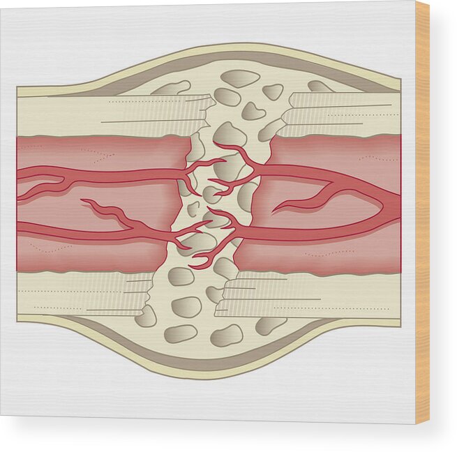
Cross Section Biomedical Illustration Of Bone Repairing Itself With New Soft Spongy Callus Developing On Framework Provided By Fibrous Tissue Joining The Broken Ends Wood Print By Dorling Kindersley from render.fineartamerica.com Table 1 describes the bone markings, which are illustrated in (figure 4). Compact bone is the outer layer and the spongy bone forms the inner layer. Now that you know what bones do, let's take a look at what they're made of and their anatomy. Marrow in the shaft of long bones is typically yellow, with red marrow in the head through the cancellous bone. I don't find it enhances the image. Bone markings the surface features of bones vary considerably, depending on the function and location in the body. Bone matrix and cells bone matrix osseous tissue is a connective tissue and like all connective tissues contains relatively few cells and large amounts of extracellular matrix. There are trabeculae in spongy bone which gives its sponge like appearance.
Related posts of cross section of human bone diagram muscles and bones of the human body.
Compact bone, spongy bone, and bone marrow. Why is the marrow red? Cross‐sectional area is derived from the integral of the bone mass profile across the narrow region. The central tubular region of the bone, called the diaphysis, flares outward near the end to form the metaphysis, which contains a largely cancellous, or spongy, interior. Huge collection, amazing choice, 100+ million high quality, affordable rf and rm images. I don't find it enhances the image. This is known as the periosteum. Diagram with articular cartilage, marrow, spongy bone, medullary cavity, endosteum, diaphysis, and periosteum. There are three general classes of bone markings: The large dark spots are passages for blood vessels and nerves. Describes the bone markings, which are illustrated in (). Bone matrix and cells bone matrix osseous tissue is a connective tissue and like all connective tissues contains relatively few cells and large amounts of extracellular matrix. While it is not as hard as compact bone, spongy bone plays an important role of protecting the marrow where blood cells are produced.
The central tubular region of the bone, called the diaphysis, flares outward near the end to form the metaphysis, which contains a largely cancellous, or spongy, interior. Human bone, cross section diagram of femur showing osteon, veins, marrow. Terms in this set (3) epiphysis. The compact bone is made up of osteon. The surface features of bones vary considerably, depending on the function and location in the body.
Bone Structure Anatomy And Physiology I from s3-us-west-2.amazonaws.com Explaned distal and proximal epiphysis. The upper (biting) surfaces of the tooth are at top, with the lower sections (bottom) embedded in the gums and jaw bone (not shown). Table 1 describes the bone markings, which are illustrated in (figure 4). Microscopic structure of compact bone consists of multiple cylindrical structural units known as osteons or haversian systems. When modeled as a beam, a long bone's cross‐sectional geometry can provide measures of its compressive strength (e.g., cross‐sectional area csa and cortical area ca), as well as its resistance to bending and torsion about a particular axis (i.e., second moments of area i and polar moments of area j , respectively; There are three general classes of bone. There are trabeculae in spongy bone which gives its sponge like appearance. It consists of two layers;
Would it be a good thing to show the epiphyseal plate?
Browse 53 bone marrow cross section stock photos and images available, or search for bone cross section or bone cells to find more great stock photos and pictures. Huge collection, amazing choice, 100+ million high quality, affordable rf and rm images. And why does the marrow stop where it does, and so sharply? After a fracture, woven bone forms initially and is gradually replaced by lamellar bone during a process known as bony substitution. This is known as the periosteum. Beautiful tooth cross section illustration, deep blue background and sparkling lights around. As the names suggest compact bone looks compact and the spongy bone looks like sponges. An outer 'fibrous layer' containing mainly fibroblasts, and an inner 'cambium layer' containing progenitor cells. I don't find it enhances the image. Internal structure of a human long bone, with a magnified cross section of the interior. Compact bone is the outer layer and the spongy bone forms the inner layer. The large dark spots are passages for blood vessels and nerves. Muscles and bones of the human body 12 photos of the muscles and bones of the human body anatomy bones of the human body quiz, major muscles and bones in the human body, muscles and bones in the human body, number of muscles and bones in the human body.
Bone matrix and cells bone matrix osseous tissue is a connective tissue and like all connective tissues contains relatively few cells and large amounts of extracellular matrix. Related posts of cross section of a long bone bone structure right foot. While it is not as hard as compact bone, spongy bone plays an important role of protecting the marrow where blood cells are produced. An outer 'fibrous layer' containing mainly fibroblasts, and an inner 'cambium layer' containing progenitor cells. Would it be a good thing to show the epiphyseal plate?
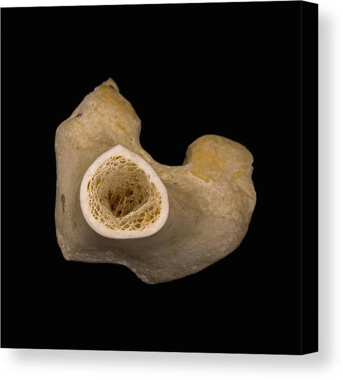
Bone Cross Section Lm Canvas Print Canvas Art By Science Stock Photography from render.fineartamerica.com The cortical bone equivalent area of the cross‐section of the region of interest (femoral neck or shaft), with all soft tissue voids (trabecular and cellular spaces) eliminated (cm 2). End of a long bone. Compact bone, spongy bone, and bone marrow. Browse 4,246 bone cross section stock photos and images available, or search for human bone cross section to find more great stock photos and pictures. Huge collection, amazing choice, 100+ million high quality, affordable rf and rm images. No need to register, buy now! Beautiful tooth cross section illustration, deep blue background and sparkling lights around. It consists of two layers;
The compact bone is made up of osteon.
There are three general classes of bone markings: Browse 53 bone marrow cross section stock photos and images available, or search for bone cross section or bone cells to find more great stock photos and pictures. Compact bone is the outer layer and the spongy bone forms the inner layer. Browse 4,246 bone cross section stock photos and images available, or search for human bone cross section to find more great stock photos and pictures. Human bone, cross section diagram of femur showing osteon, veins, marrow. Would it be a good thing to show the epiphyseal plate? Two types of bone tissues in cross section of a long bone : Internal structure of a human long bone, with a magnified cross section of the interior. Start studying cross section of bone. Huge collection, amazing choice, 100+ million high quality, affordable rf and rm images. Table 1 describes the bone markings, which are illustrated in (figure 4). The large dark spots are passages for blood vessels and nerves. After a fracture, woven bone forms initially and is gradually replaced by lamellar bone during a process known as bony substitution.

0 Comments:
Posting Komentar